


Are You Suffering From Nerve Damage?


Neuropathy is a condition that can lead to numbness or loss of sensation in the feet, but it can also cause pain. This condition is more likely to develop for those who are diabetic, obese, or with high blood pressure.
Medical Conditions Associated With Cracked Heels
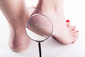
Cracked heels, a common foot condition, often arise from dry skin, which can be exacerbated by various medical conditions. For instance, individuals with diabetes may develop neuropathy, a nerve condition that impairs the ability to sweat, leading to increased skin dryness. This lack of perspiration contributes significantly to the development of dry, cracked heels. Similarly, Sjogren’s syndrome, predominantly observed in women, is an autoimmune disorder where the body mistakenly attacks its moisture-producing glands, including those responsible for perspiration. This attack results in notably dry skin, heightening the risk of cracked heels. Additionally, specific skin conditions such as psoriasis, eczema, and fungal infections directly affect the skin's moisture balance. These conditions lead to the skin becoming dry and less elastic, making it more prone to cracking, especially in the heel area, which bears significant weight and pressure during walking and standing. These medical factors underscore the importance of understanding and managing underlying health conditions to prevent or treat cracked heels effectively. If you have cracked heels, it is suggested that you schedule an appointment with a podiatrist for an evaluation and a determination of whether there is an underlying medical problem, in addition to receiving correct treatment.
If the skin on your feet starts to crack, you may want to see a podiatrist to find treatment. If you have any concerns, contact David K. Morris, DPM from Florida. Our doctor can provide the care you need to keep you pain-free and on your feet.
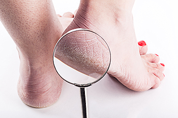
Cracked Heels
It is important to moisturize your cracked heels in order to prevent pain, bleeding, and infection. The reason cracked heels form is because the skin on the foot is too dry to support the immense pressure placed on them. When the foot expands, the dry skin on the foot begins to split.
Ways to Help Heal Them
- Invest in a good foot cream
- Try Using Petroleum Jelly
- Ease up on Soaps
- Drink Plenty of Water
Ways to Prevent Cracked Heels
- Moisturize After Showering
- Skip a Shower
- Keep Shower Water Lukewarm
- Don’t Scrub Your Feet
If you are unsure how to proceed in treating cracked heels, seek guidance from a podiatrist. Your doctor will help you with any questions or information you may need.
If you have any questions, please feel free to contact our office located in Plantation, FL . We offer the newest diagnostic and treatment technologies for all your foot care needs.
Solutions for Cracked Heels
Cracked heels may make you want to think twice about showing off your feet in warmer weather. However, cracked heels may be harmful to more than just the appearance of your feet. If deep fissures and cracks develop in your heels, they may make walking and standing painful for you. Additionally, these openings make way for germs to enter through your skin and cause infection.
There are several different causes of cracked heels. One of the most common reasons for this ailment is dry skin. This problem may make your keeps feel rough tight and itchy. Dry skin may be caused by cold air, extremely hot water, harsh soaps, and aging. Skin disorders such as eczema and psoriasis may eventually lead to dry skin. In some cases, complications may arise from cracked heels. Some of these complications are a loss of feeling in the heel, cellulitis, or a diabetic foot ulcer.
There are ways you can try to prevent getting cracked heels. One of the best ways to do so is to avoid wearing flip flops and sandals because these shoes increase your risk of drying out your feet. You should also avoid wearing shoes with a tall skinny heel, because these shoes cause your heel to expand sideways. At night, you should slather on a thick moisturizing cream on your feet and then cover them in socks to keep your feet moisturized overnight. Drinking water to stay hydrated is also a good way to ensure that your skin doesn’t become dry.
If you suffer from a severe case of cracked feet, you should make an appointment with your podiatrist to see what treatment methods are best for you.
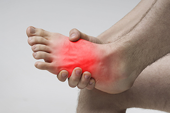
Understanding How to Live With Gout

Gout, a form of arthritis, paints a distinctive portrait of discomfort that individuals must learn to decipher. Recognizing the signs is pivotal, and sudden, intense joint pain, often in the big toe, signifies the onset. Swelling and redness can accompany these flares, underscoring the need for early identification. The causes of gout reside in an excess of uric acid, leading to the formation of sharp crystals in the joints. Factors such as diet, genetics, and underlying health conditions contribute to the heightened uric acid levels. Living with gout necessitates a multidimensional approach. Adopting a gout-friendly diet, staying hydrated, and managing weight are fundamental steps. Navigating life with gout demands an understanding of one's body and a commitment to a holistic lifestyle that fosters joint health and overall well-being. If you are afflicted with gout, it is strongly suggested that you are under the care of a podiatrist who can help you to manage this condition.
Gout is a painful condition that can be treated. If you are seeking treatment, contact David K. Morris, DPM from Florida. Our doctor will treat your foot and ankle needs.
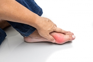
What Is Gout?
Gout is a form of arthritis that is characterized by sudden, severe attacks of pain, redness, and tenderness in the joints. The condition usually affects the joint at the base of the big toe. A gout attack can occur at any random time, such as the middle of the night while you are asleep.
Symptoms
- Intense Joint Pain - Usually around the large joint of your big toe, and it most severe within the first four to twelve hours
- Lingering Discomfort - Joint discomfort may last from a few days to a few weeks
- Inflammation and Redness -Affected joints may become swollen, tender, warm and red
- Limited Range of Motion - May experience a decrease in joint mobility
Risk Factors
- Genetics - If family members have gout, you’re more likely to have it
- Medications - Diuretic medications can raise uric acid levels
- Gender/Age - Gout is more common in men until the age of 60. It is believed that estrogen protects women until that point
- Diet - Eating red meat and shellfish increases your risk
- Alcohol - Having more than two alcoholic drinks per day increases your risk
- Obesity - Obese people are at a higher risk for gout
Prior to visiting your podiatrist to receive treatment for gout, there are a few things you should do beforehand. If you have gout you should write down your symptoms--including when they started and how often you experience them, important medical information you may have, and any questions you may have. Writing down these three things will help your podiatrist in assessing your specific situation so that he or she may provide the best route of treatment for you.
If you have any questions, please feel free to contact our office located in Plantation, FL . We offer the newest diagnostic and treatment technologies for all your foot care needs.
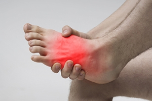
Gout
Gout is a form of arthritis that is caused by a buildup of uric acid crystals in the joints. This considered to be one of the most frequently recorded medical illnesses throughout history. Gout occurrences in the US have risen within the past twenty years and the condition now affects 8.3 million people which is 4% of all Americans. Researchers have found that gout affects men more than women and African-American men more than white men.
Symptoms of gout are warmth, swelling, discoloration, and tenderness in the affected joint area. The small joint on the big toe is the most common place for a gout attack to occur.
People who are obese, gain weight excessively, drink alcohol heavily, have high blood pressure, or have abnormal kidney function are more likely to develop gout. Furthermore, certain drugs and diseases are likely to increase levels of uric acid in the joints which eventually leads to gout. You are also more likely to develop gout if you eat a lot of meat and fish.
Many who experience gout attacks will experience repeated attacks over the years. Some people who have gout symptoms, may never have them again, but others may experience them several times a year. If you have gout symptoms throughout the year, you may have recurrent gout. Those who have gout should also be careful about their urate crystals collecting in their urinary tract, because this may lead to kidney stones.
Diagnosis for gout is done by checking the level of uric acid in the joints and blood. Your podiatrist may also prescribe medicine to reduce uric acid buildup in the blood, which will help prevent any gout attacks.
To treat gout, your podiatrist may also prescribe you Anti-inflammatory medication (NSAIDs) which will relieve the pain and swelling of a gout episode and it can also shorten a gout attack. Maintaining a healthy diet is also a proven method to prevent gout attacks.
Diagnosing an Achilles Tendon Rupture


An Achilles tendon rupture often presents suddenly, typically accompanied by a sharp pain in the back of the leg and sometimes an audible snapping sound. Many people with this injury mistakenly believe they have been hit in the calf. When examining for an Achilles rupture, the presence of swelling and bruising varies and is not entirely reliable. The gap in the tendon that indicates a rupture is often not easily palpable due to local swelling. In terms of mobility, patients can usually still perform some active movement in the ankle because other tendons also connect the calf to the foot. Pain affects passive movement but does not always limit it. Despite a ruptured Achilles, those affected can often still walk and may even be able to stand on tiptoes when using both feet, though not on the injured limb alone. The Simmonds' calf squeeze test is a reliable way for podiatrists to check for an Achilles tendon rupture. The test is done by having the patient kneel or lie prone with dangling feet. The healthy calf is used to see the foot flex, while no movement on the injured side indicates a ruptured Achilles tendon. If you believe you may have injured your Achilles tendon, it is strongly suggested that you make an appointment with a podiatrist for a thorough evaluation, diagnosis, and appropriate treatment.
Achilles tendon injuries need immediate attention to avoid future complications. If you have any concerns, contact David K. Morris, DPM of Florida. Our doctor can provide the care you need to keep you pain-free and on your feet.
What Is the Achilles Tendon?
The Achilles tendon is a tendon that connects the lower leg muscles and calf to the heel of the foot. It is the strongest tendon in the human body and is essential for making movement possible. Because this tendon is such an integral part of the body, any injuries to it can create immense difficulties and should immediately be presented to a doctor.
What Are the Symptoms of an Achilles Tendon Injury?
There are various types of injuries that can affect the Achilles tendon. The two most common injuries are Achilles tendinitis and ruptures of the tendon.
Achilles Tendinitis Symptoms
- Inflammation
- Dull to severe pain
- Increased blood flow to the tendon
- Thickening of the tendon
Rupture Symptoms
- Extreme pain and swelling in the foot
- Total immobility
Treatment and Prevention
Achilles tendon injuries are diagnosed by a thorough physical evaluation, which can include an MRI. Treatment involves rest, physical therapy, and in some cases, surgery. However, various preventative measures can be taken to avoid these injuries, such as:
- Thorough stretching of the tendon before and after exercise
- Strengthening exercises like calf raises, squats, leg curls, leg extensions, leg raises, lunges, and leg presses
If you have any questions please feel free to contact our office located in Plantation, FL . We offer the newest diagnostic tools and technology to treat your foot and ankle needs.
The Causes, Types, and Treatments of Achilles Tendon Injuries
Tendons are fibrous tissues that connect muscles with bone. The Achilles tendon is the largest tendon in the body. It connects the calf muscles at the back of the leg with the heel, and facilitates movements such as jumping, running, and walking.
Because the Achilles tendon is engaged so frequently and bears a great deal of pressure and stress throughout the day, it can become injured. Achilles tendon injuries cause the tissue to become irritated, inflamed, and swollen. Pain can come on gradually or be immediate, and will vary from mild to severe depending upon the injury. Where the pain occurs will vary as well, from just above the heel up through the back of the leg. There may also be stiffness in the tendon.
Achilles tendon injuries can often be caused by repetitive stress. They may also occur while running, playing tennis, gymnastics, football, basketball, dancing, soccer, baseball or other sports that require speeding up, slowing down, or pivoting quickly. Wearing high heels, falling from an elevation, stepping in a hole, having flat feet, bone spurs, tight leg muscles or tendons, wearing improper athletic shoes, exercising on uneven surfaces, or starting a new type of exercise can also cause Achilles tendon injuries.
The two most common Achilles tendon injuries are tendonitis and ruptures. Tendonitis causes painful inflammation and can occur in different parts of the tendon. Non-insertional Achilles tendonitis occurs when the fibers in middle of the tendon begin to break down, thicken, and swell. This condition typically affects younger, more active adults. Insertional Achilles tendonitis occurs where the tendon inserts into the heel bone. It is common for bone spurs to form with this type of injury. This condition can affect people of any age and level of activity.
Achilles tendon ruptures are a tear in the tendon. These breaks may be partial or complete. There may be an audible popping noise at the moment of injury and the pain will be sudden and severe.
An Achilles tendon injury can be diagnosed by your podiatrist after they examine you, check your range of motion, and possibly perform a calf squeeze test or review an X-ray or MRI. Depending on the type and severity of your injury, your podiatrist may treat your condition with rest/ice/compression/elevation (RICE), nonsteroidal anti-inflammatory medications, heel lifts, and stretching and strengthening exercises. If you have torn your Achilles tendon, treatment may include physical therapy, ultrasound, shockwave therapy, or possibly even surgery.
Risk Factors for Foot Stress Fractures
Stress fractures, which are generally attributed to overuse, primarily affect weight-bearing bones and are often triggered by a sudden increase in physical activity. This serves as a significant risk factor for microscopic bone fractures in the feet. Embracing a new sport or intensifying exercise routines can elevate the likelihood of stress fractures. Biomechanical issues, such as abnormal gait patterns like flat feet or high arches and poor running form can also heighten the risk. Wearing inadequate footwear, including ill-fitting or worn-out shoes that offer insufficient support, places added stress on the foot bones. Nutritional deficiencies, particularly in calcium and vitamin D intake, weaken bones and make them more susceptible to such fractures. Hormonal changes, especially in women, like amenorrhea, can disrupt hormone balance and reduce bone density, which further increases the risk of stress fractures. Pain that starts during physical activity and worsens with continued exertion is a common symptom. Swelling in the affected area indicates inflammation, and tenderness of the bone to the slightest touch is another indicator. If these symptoms persist for about a week, it is suggested that you make an appointment with a podiatrist for a full exam, which may include imaging tests, as a foot stress fracture may not be visible on an X-ray.
Stress fractures occur when there is a tiny crack within a bone. To learn more, contact David K. Morris, DPM from Florida. Our doctor can provide the care you need to keep you pain free and on your feet.
How Are They Caused?
Stress fractures are the result of repetitive force being placed on the bone. Since the lower leg and feet often carry most of the body’s weight, stress fractures are likely to occur in these areas. If you rush into a new exercise, you are more likely to develop a stress fracture since you are starting too much, too soon. Pain resulting from stress fractures may go unnoticed at first, however it may start to worsen over time.
Risk Factors
- Gender – They are more commonly found in women compared to men.
- Foot Problems – People with unusual arches in their feet are more likely to develop stress fractures.
- Certain Sports – Dancers, gymnasts, tennis players, runners, and basketball players are more likely to develop stress fractures.
- Lack of Nutrients – A lack of vitamin D and calcium may weaken the bones and make you more prone to stress fractures
- Weak Bones – Osteoporosis can weaken the bones therefore resulting in stress fractures
Stress fractures do not always heal properly, so it is important that you seek help from a podiatrist if you suspect you may have one. Ignoring your stress fracture may cause it to worsen, and you may develop chronic pain as well as additional fractures.
If you have any questions, please feel free to contact our office located in Plantation, FL . We offer the newest diagnostic and treatment technologies for all your foot care needs.
Dealing with Stress Fractures of the Foot and Ankle
Stress fractures are small breaks in the bone that are caused by repetitive stress. They typically occur due to overuse, forcing the bones of the foot or ankle to continually absorb the full impact of each step taken. Stress fractures can also be caused by abnormal foot structure, osteoporosis, bone deformities, or wearing improper footwear during exercise.
Stress fractures are common for individuals whose daily activities cause high levels of impact on their feet and ankles. Those who run, play tennis or basketball, or practice gymnastics tend to experience these fractures more frequently. Anyone is susceptible to this problem, though. Individuals who are normally sedentary and suddenly begin an intense, high impact workout may sustain stress fractures. This is because their muscles are not yet strong enough to handle and cushion the intensity of their activity. Osteoporosis may also cause someone to get stress fractures, because the disease weakens an afflicted person's bones and makes it easier for them to break down.
Pain from stress fractures typically occurs in the general area of the fracture. Pain can also manifest as “pinpoint pain” or pain that is felt when the site of the injury is touched, and can be accompanied by swelling. It may occur during or after activity, and it may disappear while resting and return when standing or moving. Engaging in any kind of activity, high impact or otherwise, will aggravate the pain. If the intensity of the activity increases before the stress fracture has properly healed, it can cause a full fracture.
Treatment can vary depending on the individual and the degree of injury. The primary way to treat a stress fracture is to rest the hurt foot. Some fractures will heal quickly with only a little bit of rest, while others may require a long rest period and the use of crutches, immobilization, or physical therapy. Under certain circumstances, surgery may be required to install support pins around the fracture to assist in healing.
If you are undergoing a new exercise regimen in running or some other kind of high impact activity, set incremental goals on a weekly basis so you can build up muscle strength. Make sure to wear supportive shoes to better protect you feet.
If you begin to experience any symptoms of stress fractures, you should stop exercising and rest. If the symptoms persist, consult with your podiatrist. Remembering these tips can help you prevent stress fractures to your foot and ankle, and allow you to continue living normally.
Methods to Manage Foot Arthritis Pain
Foot arthritis is not only extremely painful but it also can seriously affect your ability to conduct normal daily activities. Medication can play a vital role in the relieving pain and inflammation of arthritic foot pain. Nonsteroidal anti-inflammatory drugs, or NSAIDs, help to reduce inflammation and pain, and some topical versions in the form of gels or creams can offer targeted relief. Corticosteroids swiftly control inflammation, with oral corticosteroids used for systemic issues and joint-specific injections for localized inflammation. Analgesics, including acetaminophen and opioids, focus solely on pain relief, but opioids are for short-term use due to their potential for dependency. Certain topical treatments can alleviate muscle and surface-level soft tissue pain. To help reduce the progression of arthritic foot pain, disease modifying antirheumatic drugs, or DMARDs, may be used with small joint conditions like rheumatoid arthritis. Gout medications manage uric acid levels and provide relief during flare ups. Biologics are systemic agents that alter the course of inflammatory diseases that affect various joints, including the feet. Osteoporosis medications, while not foot-specific, promote overall bone strength, and may help to reduce the risk of fractures. If you suffer from any type of arthritis that affects your feet, it is suggested that you make an appointment with a podiatrist who can recommend the appropriate medication for you.
Arthritis can be a difficult condition to live with. If you are seeking treatment, contact David K. Morris, DPM from Florida. Our doctor can provide the care you need to keep you pain-free and on your feet.
Arthritic Foot Care
Arthritis is a term that is commonly used to describe joint pain. The condition itself can occur to anyone of any age, race, or gender, and there are over 100 types of it. Nevertheless, arthritis is more commonly found in women compared to men, and it is also more prevalent in those who are overweight. The causes of arthritis vary depending on which type of arthritis you have. Osteoarthritis for example, is often caused by injury, while rheumatoid arthritis is caused by a misdirected immune system.
Symptoms
- Swelling
- Pain
- Stiffness
- Decreased Range of Motion
Arthritic symptoms range in severity, and they may come and go. Some symptoms stay the same for several years but could potentially get worse with time. Severe cases of arthritis can prevent its sufferers from performing daily activities and make walking difficult.
Risk Factors
- Occupation – Occupations requiring repetitive knee movements have been linked to osteoarthritis
- Obesity – Excess weight can contribute to osteoarthritis development
- Infection – Microbial agents can infect the joints and trigger arthritis
- Joint Injuries – Damage to joints may lead to osteoarthritis
- Age – Risk increases with age
- Gender –Most types are more common in women
- Genetics – Arthritis can be hereditary
If you suspect your arthritis is affecting your feet, it is crucial that you see a podiatrist immediately. Your doctor will be able to address your specific case and help you decide which treatment method is best for you.
If you have any questions, please feel free to contact our office located in Plantation, FL . We offer the newest diagnostic and treatment technologies for all your foot care needs.
